

ما هي الجراحة بالمنظار؟
الجراحة بالمنظار، والتي تُعرف أيضًا باسم الجراحة التنظيرية أو جراحة المنظار أو جراحة مناظير، هي إحدى أحدث التقنيات الجراحية التي أحدثت نقلة نوعية في عالم الجراحة الحديثة. وتعتمد هذه التقنية على إجراء العملية من خلال عدة فتحات صغيرة في الجلد بدلًا من الشق الجراحي الكبير المستخدم في الجراحة المفتوحة التقليدية، مما يقلل من إصابة الأنسجة ويساعد على تحسين نتائج العلاج وتسريع التعافي. وتُستخدم الجراحة بالمنظار اليوم في العديد من التخصصات الطبية، مثل الجراحة العامة، وجراحات السمنة، وجراحة النساء والتوليد، وجراحة المسالك البولية، وجراحات القولون والمستقيم، وأصبحت الخيار الأول في العديد من عمليات المنظار لما توفره من دقة وأمان ومضاعفات أقل مقارنة بالجراحة التقليدية.
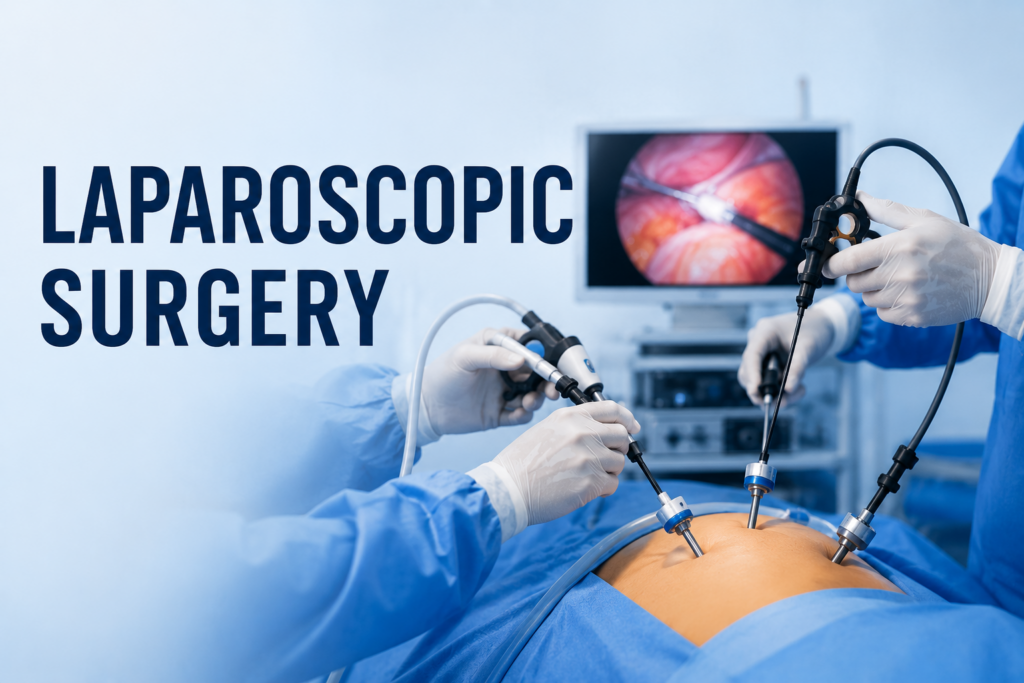
يعتمد نجاح عملية المنظار على استخدام المنظار الجراحي، وهو أنبوب رفيع وطويل مزود بكاميرا عالية الدقة ومصدر إضاءة قوي يعتمد على الألياف الضوئية. ويتم إدخال جهاز المنظار عبر فتحة صغيرة يتراوح قطرها عادة بين 5 و12 ملليمترًا، لينقل صورًا مكبرة وفورية للأعضاء الداخلية إلى شاشة عرض عالية الوضوح داخل غرفة العمليات، مما يمنح الجراح رؤية دقيقة ومفصلة لمكان الجراحة، وتوفر هذه الرؤية المكبرة للجراح القدرة على إجراء العمليات الدقيقة بكفاءة عالية مع الحفاظ على الأنسجة السليمة المحيطة وتقليل فرص حدوث إصابات غير ضرورية.
وعلى عكس الجراحة المفتوحة، التي تتطلب شقًا جراحيًا كبيرًا للوصول إلى العضو المستهدف، تعتمد الجراحة بالمنظار على مجموعة من الأدوات الجراحية الطويلة التي يتم إدخالها من خلال فتحات صغيرة باستخدام أدوات خاصة تُعرف باسم التروكارات، وتساعد هذه الأدوات الجراح على فصل الأنسجة، والسيطرة على النزيف، واستئصال الأعضاء المصابة، وإجراء مختلف الخطوات الجراحية بدقة أثناء متابعة العملية عبر شاشة العرض.
ويُعد مصطلح الجراحة التنظيرية مصطلحًا عامًا يشمل مجموعة واسعة من التقنيات الجراحية التي تهدف إلى تقليل التدخل الجراحي والحد من إصابة الأنسجة. وتُعتبر الجراحة بالمنظار أكثر هذه التقنيات انتشارًا واستخدامًا، بينما تُعد الجراحة الروبوتية وغيرها من التقنيات الموجهة بالصور امتدادًا متطورًا لنفس المفهوم، وهو تحقيق أفضل النتائج الجراحية الممكنة مع تقليل الألم والمضاعفات وتسريع تعافي المريض.
وخلال العقود الثلاثة الماضية، أصبحت الجراحة بالمنظار الخيار المفضل لإجراء العديد من العمليات داخل البطن والحوض، وذلك لما توفره من مزايا سريرية عديدة، تشمل تقليل الألم بعد الجراحة، وانخفاض فقدان الدم، وتقليل احتمالية الإصابة بعدوى الجروح، وقِصر مدة الإقامة في المستشفى، وسرعة العودة إلى الأنشطة اليومية، بالإضافة إلى الحصول على نتائج تجميلية أفضل بفضل صِغر حجم الفتحات الجراحية
وقد ساهمت هذه المزايا في اعتماد جراحات المنظار كمعيار علاجي أساسي في عدد كبير من العمليات الاختيارية وعمليات الطوارئ على حد سواء، واليوم، تواصل التطورات المستمرة في أنظمة التصوير عالية الدقة، والأدوات الجراحية المتقدمة، وأجهزة الطاقة الحديثة توسيع نطاق استخدام الجراحة بالمنظار، مما يتيح للجراحين إجراء عمليات أكثر تعقيدًا بدرجة أعلى من الدقة والأمان والكفاءة.
كيف تتم الجراحة بالمنظار؟
تعتمد الجراحة بالمنظار على تقنيات تصوير متطورة وأدوات جراحية متخصصة تسمح للجراح بإجراء العملية داخل الجسم دون الحاجة إلى عمل شق جراحي كبير. وبدلًا من رؤية الأعضاء بشكل مباشر كما هو الحال في الجراحة المفتوحة، تتم جميع خطوات عملية المنظار أثناء مشاهدة صورة مكبرة وعالية الدقة تظهر على شاشة مخصصة داخل غرفة العمليات، وهو ما يمنح الجراح رؤية أوضح ودقة أكبر مع تقليل إصابة الأنسجة المحيطة.
تبدأ العملية التنظيرية بإخضاع المريض للتخدير الكلي، لضمان الراحة التامة وعدم الشعور بأي ألم طوال فترة الجراحة. وبعد تجهيز المريض وتعقيم منطقة العملية، يقوم الجراح بإجراء فتحة واحدة أو عدة فتحات صغيرة في الجلد، يتراوح طول كل منها عادة بين 5 و12 ملليمترًا، لتكون منافذ لإدخال المنظار الطبي والأدوات الجراحية المختلفة.
ولتوفير مساحة عمل مناسبة داخل التجويف البطني، يتم ضخ غاز ثاني أكسيد الكربون (CO₂) بلطف من خلال أحد التروكارات باستخدام جهاز مخصص لذلك. وتُعرف هذه الخطوة باسم Pneumoperitoneum، حيث يرتفع جدار البطن بعيدًا عن الأعضاء الداخلية، مما يمنح الجراح رؤية واضحة ومساحة كافية لتحريك الأدوات الجراحية بأمان.
بعد ذلك، يتم إدخال المنظار الجراحي المزود بكاميرا عالية الدقة ومصدر إضاءة بالألياف الضوئية عبر أحد المنافذ الجراحية. وتنقل الكاميرا صورًا مكبرة وفورية للأعضاء الداخلية إلى شاشات العرض داخل غرفة العمليات، مما يسمح للفريق الجراحي برؤية التفاصيل التشريحية بدقة فائقة، وغالبًا ما تكون هذه الرؤية أوضح من الرؤية المباشرة في الجراحة المفتوحة التقليدية. وبعد تثبيت جهاز المنظار، يتم إدخال باقي الأدوات الجراحية من خلال منافذ إضافية حسب نوع العملية، وتشمل هذه الأدوات الملاقط، وأدوات فصل الأنسجة، والمقصات، وحامل الإبر، وأجهزة الشفط والغسيل، وأدوات إبعاد الأنسجة.
وفي العمليات الأكثر تعقيدًا، قد يستخدم الجراح أجهزة طاقة متقدمة تساعد على قطع الأنسجة وإغلاق الأوعية الدموية في الوقت نفسه، مثل Harmonic Scalpel وLigaSure وThunderbeat، والتي ساهمت بشكل كبير في تحسين كفاءة جراحات المنظار من خلال تقليل النزيف، وتقصير مدة العملية، والحد من انتقال الحرارة إلى الأنسجة المجاورة. كما أظهرت الدراسات المقارنة أن لكل نظام من هذه الأنظمة مميزات تختلف باختلاف نوع العملية والأهداف الجراحية.
وبعد الانتهاء من جميع الخطوات الجراحية، يقوم الجراح بفحص مكان العملية بعناية للتأكد من السيطرة الكاملة على النزيف وعدم وجود أي مضاعفات. وإذا تطلبت العملية استئصال عضو أو عينة نسيجية، يتم إخراجها من خلال أحد المنافذ الجراحية، وقد يتم توسيع إحدى الفتحات قليلًا عند الحاجة.
وفي نهاية عملية المناظير، يتم تفريغ غاز ثاني أكسيد الكربون من التجويف البطني، ثم إزالة جميع الأدوات والمناظير وإغلاق الفتحات الصغيرة باستخدام الغرز الجراحية أو اللاصق الطبي أو الشرائط الجراحية المعقمة. ونظرًا لصغر حجم هذه الفتحات مقارنة بالجراحة المفتوحة، فإن المرضى غالبًا ما يشعرون بألم أقل بعد العملية، ويتعافون بصورة أسرع، مع بقاء آثار جراحية بسيطة للغاية.
تاريخ الجراحة بالمنظار
يمتد تاريخ الجراحة بالمنظار لأكثر من قرن من الزمان، ويعكس رحلة طويلة من التطور والابتكار في مجال العلوم الجراحية والتكنولوجيا الطبية. فما بدأ في البداية كوسيلة لتشخيص أمراض وتجويف البطن، تطور تدريجيًا ليصبح أحد أهم الإنجازات في الجراحة التنظيرية الحديثة، مُحدثًا تغييرًا جذريًا في طريقة إجراء العمليات الجراحية بمختلف التخصصات الطبية.
تعود البدايات الأولى لاستخدام المنظار الطبي إلى أوائل القرن العشرين، عندما بدأ الأطباء في استكشاف وسائل لفحص التجويف البطني باستخدام أجهزة المناظير. إلا أن الإمكانيات التقنية في ذلك الوقت كانت محدودة، حيث كانت الإضاءة ضعيفة، والعدسات البصرية بدائية، كما لم تكن الأدوات الجراحية المتخصصة متوفرة، وهو ما جعل استخدام المناظير يقتصر في البداية على التشخيص فقط، دون إجراء تدخلات علاجية.
وخلال النصف الثاني من القرن العشرين، شهدت الجراحة بالمنظار تطورًا كبيرًا بفضل ظهور أنظمة الإضاءة المعتمدة على الألياف الضوئية، وتحسن جودة العدسات، وابتكار الكاميرات المصغرة. وقد ساهمت هذه التطورات في تحسين جودة الصورة بشكل كبير، ومنحت الجراحين رؤية أكثر وضوحًا للتراكيب التشريحية داخل الجسم، مما مهد الطريق لتحول عملية التنظير من وسيلة تشخيصية إلى تقنية جراحية متكاملة تُستخدم لعلاج العديد من الأمراض.
وجاءت نقطة التحول الحقيقية في عام 1987 عندما نجح الجراح الفرنسي Philippe Mouret في إجراء أول استئصال للمرارة بالمنظار. وقد أثبت هذا الإنجاز أن العمليات الجراحية المعقدة يمكن إجراؤها من خلال فتحات صغيرة مع تحقيق نتائج ممتازة، وتقليل الألم بعد الجراحة، وتسريع فترة التعافي مقارنة بالجراحة المفتوحة، وهو ما أدى إلى انتشار جراحات المنظار في مختلف أنحاء العالم.
وخلال تسعينيات القرن الماضي، ساهم التطور السريع في أنظمة التصوير، وجهاز المنظار، وأدوات الجراحة، وأجهزة ضخ الغاز، بالإضافة إلى التدريب المتقدم للجراحين، في توسيع نطاق استخدام الجراحة بالمنظار عالميًا. وأصبحت العديد من عمليات المنظار، مثل استئصال الزائدة الدودية، وإصلاح الفتق، وجراحات القولون والمستقيم، وجراحات السمنة، وجراحات النساء، وجراحات المسالك البولية، تُجرى بشكل روتيني باستخدام هذه التقنية.
وفي القرن الحادي والعشرين، شهدت الجراحة التنظيرية مرحلة جديدة من التطور مع ظهور أنظمة التصوير عالية الدقة (HD) وتقنيات 4K، والتصوير ثلاثي الأبعاد، والجراحة الموجهة بالفلورسنت، بالإضافة إلى أجهزة الطاقة المتقدمة القادرة على قطع الأنسجة وإغلاق الأوعية الدموية في الوقت نفسه. كما ساهمت الدراسات المقارنة بين تقنيات مثل Thunderbeat وLigaSure وHarmonic Scalpel في تحسين أداء جراحة المنظار من خلال زيادة الدقة، وتقليل فقدان الدم، ورفع كفاءة العديد من الإجراءات الجراحية.
واليوم، تُعد الجراحة بالمنظار أحد الركائز الأساسية في الجراحة التنظيرية الحديثة، ولا تزال تشهد تطورًا مستمرًا بفضل تقنيات الجراحة الروبوتية، والذكاء الاصطناعي، وأنظمة الملاحة الجراحية، والتقنيات الرقمية المتقدمة. ومن المتوقع أن تواصل عمليات المنظار تطورها خلال السنوات القادمة لتوفر مستويات أعلى من الدقة والأمان وتحسين نتائج علاج المرضى في مختلف التخصصات الطبية.
مكونات نظام الجراحة بالمنظار
يعتمد نجاح الجراحة بالمنظار بشكل كبير على تكامل خبرة الجراح مع منظومة متطورة من الأجهزة والأدوات الجراحية المتخصصة. ويجمع نظام جراحة المنظار الحديث بين تقنيات التصوير المتقدمة، والأدوات الجراحية الدقيقة، وأجهزة الطاقة الحديثة، مما يوفر رؤية واضحة وتحكمًا دقيقًا أثناء إجراء العملية، ويساهم في تحقيق أعلى مستويات الأمان والكفاءة.
المنظار الجراحي (Laparoscope)
يُعد المنظار الجراحي المكون الأساسي في الجراحة بالمنظار، وهو عبارة عن أنبوب رفيع وطويل مزود بعدسات بصرية عالية الجودة تنقل صورًا دقيقة من داخل الجسم. وتتوفر المناظير بأحجام وزوايا رؤية مختلفة، بما يسمح للجراح باختيار النوع المناسب وفقًا لطبيعة العملية.
نظام الكاميرا (Camera System)
تُثبت كاميرا رقمية عالية الدقة على المنظار الجراحي لنقل صور مباشرة إلى شاشات غرفة العمليات. وقد ساهمت تقنيات التصوير الحديثة، مثل Full HD و4K والتصوير ثلاثي الأبعاد، في تحسين إدراك العمق ودقة التعرف على التراكيب التشريحية أثناء عمليات المنظار.
مصدر الإضاءة (Light Source)
نظرًا لأن التجويف البطني معتم بطبيعته، يعتمد جهاز المنظار على مصدر إضاءة قوي يعمل بالألياف الضوئية أو بتقنية LED لتوفير رؤية واضحة أثناء الجراحة، وهو ما يساعد الجراح على تمييز الأنسجة والأوعية الدموية والهياكل التشريحية المهمة بدقة.
شاشة العرض (Video Monitor)
تُعرض الصور التي يلتقطها المنظار الطبي على شاشة عالية الدقة داخل غرفة العمليات، حيث يُجري الجراح جميع خطوات الجراحة بالمنظار من خلال متابعة هذه الصور المكبرة بدلًا من النظر مباشرة إلى المريض، مما يوفر دقة أعلى أثناء التعامل مع الأنسجة الدقيقة.
جهاز ضخ ثاني أكسيد الكربون (CO₂ Insufflation System)
يقوم هذا الجهاز بضخ غاز ثاني أكسيد الكربون داخل التجويف البطني لتكوين مساحة عمل مناسبة تُعرف باسم Pneumoperitoneum، مما يحسن الرؤية، ويسهل حركة الأدوات الجراحية، ويساعد على حماية الأعضاء الداخلية أثناء عملية المنظار.
التروكارات والقنيات (Trocars and Cannulas)
تُستخدم التروكارات لإنشاء منافذ صغيرة في جدار البطن يتم من خلالها إدخال المنظار الجراحي وبقية الأدوات الجراحية. وتختلف أحجامها بحسب نوع العملية، ويتراوح قطرها غالبًا بين 5 و12 ملليمترًا.
الأدوات الجراحية الخاصة بالمنظار
تتميز أدوات جراحة المنظار بأنها طويلة ورفيعة لتناسب العمل من خلال الفتحات الصغيرة. وتشمل الملاقط، وأدوات فصل الأنسجة، والمقصات، وحوامل الإبر، وأجهزة الشفط والغسيل، وأدوات الإبعاد، والتي تتيح للجراح تنفيذ مختلف خطوات العملية بدقة مع تقليل إصابة الأنسجة.
أجهزة الطاقة (Energy Devices)
تعتمد الجراحة بالمنظار الحديثة على أجهزة طاقة متطورة لقطع الأنسجة وإغلاق الأوعية الدموية مع تقليل النزيف والانتشار الحراري. ومن أشهر هذه التقنيات:
- الجراحة الكهربائية أحادية القطب (Monopolar Electrosurgery)
- الجراحة الكهربائية ثنائية القطب (Bipolar Electrosurgery)
- LigaSure™ Vessel Sealing System
- Harmonic Scalpel
- Thunderbeat
وقد أثبتت الدراسات أن لكل جهاز خصائص مختلفة فيما يتعلق بكفاءة إغلاق الأوعية الدموية، والانتشار الحراري، وسرعة الأداء، ودقة فصل الأنسجة، ويعتمد اختيار التقنية المناسبة على نوع العملية، وطبيعة الأنسجة، وخبرة الجراح، والأهداف العلاجية.
أنواع الجراحة بالمنظار
شهدت الجراحة بالمنظار تطورًا كبيرًا خلال العقود الماضية، وأصبحت تضم مجموعة متنوعة من التقنيات التي تلبي الاحتياجات التشخيصية والعلاجية المختلفة. ويعتمد اختيار نوع عملية المنظار على حالة المريض، وطبيعة المرض، ومدى تعقيد الإجراء الجراحي، بالإضافة إلى خبرة الجراح.
المنظار التشخيصي (Diagnostic Laparoscopy)
يُستخدم المنظار التشخيصي لفحص التجويف البطني أو الحوض بشكل مباشر عندما لا تكفي الفحوصات أو الأشعة للوصول إلى تشخيص دقيق. ويُستخدم في تقييم آلام البطن غير المبررة، والعقم، وأمراض الكبد، والانتباذ البطاني الرحمي، وإصابات البطن، وبعض الأورام، مع إمكانية أخذ عينات نسيجية عند الحاجة.
الجراحة العلاجية بالمنظار (Operative Laparoscopy)
تشمل الجراحة العلاجية بالمنظار إجراء العمليات الجراحية الكاملة باستخدام التقنيات التنظيرية، وأصبحت الخيار القياسي للعديد من عمليات المنظار مثل استئصال المرارة، واستئصال الزائدة الدودية، وإصلاح الفتق، وجراحات السمنة، وجراحات القولون والمستقيم، وجراحات النساء.
جراحة المناظير المتقدمةالجراحة المتقدمة بالمنظار (Advanced Laparoscopic Surgery)
تشمل جراحات المنظار المتقدمة العمليات المعقدة التي تتطلب تدريبًا عاليًا وخبرة كبيرة، مثل تحويل مسار المعدة، واستئصال أجزاء من القولون، وجراحات البنكرياس والكبد، واستئصال الغدة الكظرية، وإصلاح ارتجاع المريء.
الجراحة بالمنظار عبر فتحة واحدة (Single-Incision Laparoscopic Surgery - SILS)
تعتمد هذه التقنية على إجراء العملية من خلال فتحة واحدة فقط، غالبًا داخل السرة، بهدف تقليل عدد الشقوق الجراحية وتحسين النتيجة التجميلية، مع الحفاظ على مبادئ الجراحة التنظيرية، رغم أنها تتطلب مهارة جراحية أكبر.
الجراحة الروبوتية بالمنظار (Robotic-Assisted Laparoscopic Surgery)
تُعد الجراحة الروبوتية أحد التطورات الحديثة في الجراحة بالمنظار، حيث يستخدم الجراح نظامًا روبوتيًا للتحكم في الأدوات الجراحية بدقة عالية. ويوفر هذا النظام مدى حركة أكبر، ورؤية ثلاثية الأبعاد، وتقليل الاهتزاز، مما يجعله مناسبًا لبعض العمليات المعقدة، مع بقاء الجراح هو المتحكم الكامل في جميع خطوات العملية.
أشهر العمليات التي تُجرى باستخدام الجراحة بالمنظار
أصبحت الجراحة بالمنظار الخيار الأول لإجراء العديد من العمليات الجراحية في مختلف التخصصات الطبية، وذلك بفضل التطور المستمر في أجهزة التصوير، والأدوات الجراحية، وتقنيات الطاقة الحديثة. وقد ساهمت هذه التطورات في توسيع نطاق عمليات المنظار لتشمل إجراءات كانت تُجرى سابقًا بالجراحة المفتوحة، مع الحفاظ على نفس الكفاءة العلاجية وتقليل فترة التعافي.
Common laparoscopic procedures include:
الجراحة العامة
- Laparoscopic cholecystectomy (gallbladder removal)
- Laparoscopic appendectomy
- Laparoscopic inguinal and ventral hernia repair
- Splenectomy
- Adrenalectomy
جراحات السمنة
- Laparoscopic sleeve gastrectomy
- Laparoscopic Roux-en-Y gastric bypass
- One Anastomosis Gastric Bypass (OAGB)
- عملية السادي إس
- Revisional bariatric surgery
Colorectal Surgery
- Colectomy
- Sigmoidectomy
- Low anterior resection
- Rectal cancer surgery
Upper Gastrointestinal Surgery
- جراحات فتق الحجاب الحاجز بالمنظار
- Fundoplication for gastroesophageal reflux disease (GERD)
- Gastric tumor resection
Gynecologic Surgery
- Hysterectomy
- Ovarian cystectomy
- Endometriosis surgery
- Treatment of ectopic pregnancy
- Myomectomy
Urologic Surgery
- Nephrectomy
- Pyeloplasty
- Prostate surgery
- Adrenal surgery
As technology continues to advance, increasingly complex procedures are being performed laparoscopically, providing patients with the benefits of minimally invasive surgery without compromising surgical effectiveness.
Advantages of Laparoscopic Surgery
The widespread adoption of laparoscopic surgery has been driven by its ability to achieve outcomes comparable to, and in many cases superior to, traditional open surgery while significantly reducing surgical trauma. Numerous clinical studies have demonstrated that minimally invasive techniques improve both short-term recovery and overall patient experience.
Compared with open surgery, laparoscopic surgery offers several important advantages:
- Smaller skin incisions and improved cosmetic outcomes
- Reduced postoperative pain and discomfort
- Less blood loss during surgery
- Lower risk of wound infection
- Reduced tissue trauma
- Shorter hospital stay
- Faster return to normal daily activities and work
- Earlier recovery of bowel function
- Lower incidence of postoperative adhesions in selected procedures
- Improved visualization through magnified high-definition imaging
- Reduced need for postoperative pain medication
- Enhanced patient satisfaction and quality of recovery
For these reasons, laparoscopic surgery has become the preferred surgical approach whenever it is technically feasible and clinically appropriate.
Limitations and Challenges
Despite its many advantages, laparoscopic surgery is not suitable for every patient or every clinical situation. Successful outcomes depend on careful patient selection, appropriate surgical planning, and the experience of the operating team.
Several limitations should be considered:
- A significant learning curve is required for surgeons to develop advanced laparoscopic skills.
- Specialized equipment and operating room infrastructure increase procedural costs.
- Reduced tactile feedback compared with open surgery may make certain dissections more challenging.
- Complex anatomy, extensive adhesions, or severe inflammation may limit the feasibility of a laparoscopic approach.
- Unexpected bleeding or intraoperative complications may require conversion to open surgery to ensure patient safety.
- Equipment malfunction or limited instrument mobility can occasionally complicate technically demanding procedures.
- Certain emergencies or advanced malignancies may still require conventional open surgery.
Although these challenges exist, continuous improvements in surgical training, imaging technology, and instrument design have steadily expanded the range of conditions that can be managed laparoscopically.
Risks and Possible Complications
Like all surgical procedures, laparoscopic surgery carries potential risks and complications. However, when performed by experienced surgeons using appropriate patient selection and modern surgical techniques, serious complications remain relatively uncommon.
Potential complications include:
النزيف.
Bleeding may occur from injured blood vessels during trocar insertion or tissue dissection. Most cases can be controlled laparoscopically, although severe bleeding may require conversion to open surgery.
العدوى.
Although the risk of surgical site infection is lower than in open surgery, wound infections and intra-abdominal infections can still occur.
Injury to Internal Organs
Adjacent organs such as the bowel, bladder, liver, spleen, ureters, or major blood vessels may rarely be injured during trocar placement or surgical dissection.
Carbon Dioxide – Related Complications
Because laparoscopic surgery requires insufflation of carbon dioxide, some patients may experience temporary shoulder pain, abdominal discomfort, or gas retention after surgery. Rare complications such as gas embolism or subcutaneous emphysema have also been reported.
Port-Site Hernia
Although uncommon, a hernia may develop at the site where larger trocars are inserted, particularly if the abdominal wall is not adequately closed.
Blood Clots
As with any major operation, there is a small risk of developing deep vein thrombosis (DVT) or pulmonary embolism, particularly in high-risk patients. Early mobilization and preventive measures significantly reduce this risk.
Anesthesia-Related Risks
General anesthesia may be associated with temporary nausea, vomiting, allergic reactions, respiratory complications, or cardiovascular events, depending on the patient’s overall health status.
Overall, laparoscopic surgery has an excellent safety profile, and for appropriately selected patients, the benefits of minimally invasive surgery generally outweigh the potential risks. Careful preoperative evaluation, adherence to established surgical protocols, and experienced surgical teams are essential for minimizing complications and achieving the best possible outcomes.
Who Is a Candidate for Laparoscopic Surgery?
Laparoscopic surgery is an excellent option for many patients requiring abdominal or pelvic surgery. However, eligibility depends on several factors, including the patient’s overall health, the underlying condition, previous surgical history, and the complexity of the procedure. The final decision is made after a thorough medical evaluation by the surgeon, Patients who may be suitable candidates for laparoscopic surgery include those undergoing:
- Gallbladder removal (cholecystectomy)
- Appendectomy
- Hernia repair
- جراحة السمنة
- Colorectal surgery
- Anti-reflux surgery
- Certain gynecologic and urologic procedures
While most patients benefit from a minimally invasive surgery approach, factors such as extensive scar tissue, severe infection, uncontrolled bleeding, or advanced disease may influence the choice of surgical technique.
When Is Open Surgery Preferred?
Although laparoscopic surgery is considered the preferred approach for many procedures, open surgery remains the safest and most appropriate option in certain situations. A surgeon may recommend open surgery when:
- The disease is highly advanced or involves multiple organs.
- Extensive scar tissue (adhesions) from previous surgeries limits laparoscopic access.
- Severe inflammation or infection obscures normal anatomy.
- Significant uncontrolled bleeding occurs during surgery.
- Large tumors require direct access for safe removal.
- Emergencies demand rapid surgical intervention.
- Patient-specific factors make minimally invasive surgery unsafe.
In some cases, a procedure may begin laparoscopically but be converted to open surgery if necessary to ensure the best possible outcome and patient safety.
Laparoscopic Surgery vs Open Surgery
Both laparoscopic surgery and open surgery are effective surgical approaches, and the best choice depends on the patient’s condition, the complexity of the procedure, and the surgeon’s clinical judgment. While laparoscopic surgery has become the preferred option for many operations due to its minimally invasive nature, open surgery remains essential in selected complex or emergency cases.
Laparoscopic Surgery
- Performed through several small incisions.
- Uses a high-definition camera and specialized instruments.
- Causes less postoperative pain.
- Typically results in less blood loss.
- Offers a shorter hospital stay.
- Allows faster recovery and return to daily activities.
- Leaves smaller, less noticeable scars.
- Generally has a lower risk of wound infection.
Open Surgery
- Requires a larger incision to access the surgical area directly.
- Provides direct visualization of organs and tissues.
- May be preferred for highly complex procedures or emergency situations.
- Can be more suitable when extensive adhesions, large tumors, or severe bleeding are present.
- Usually involves a longer recovery period and a higher level of postoperative discomfort.
Ultimately, the decision between laparoscopic surgery and open surgery should be individualized, balancing the patient’s condition, surgical goals, and overall safety.
Laparoscopic Surgery vs Robotic Surgery
Both laparoscopic surgery and robotic-assisted surgery are forms of minimally invasive surgery designed to reduce tissue trauma and promote faster recovery. The main difference lies in the technology used to perform the operation.
Laparoscopic Surgery
- The surgeon directly controls the laparoscopic instruments by hand.
- Uses a high-definition camera to visualize the surgical field.
- Proven safe and effective for a wide range of procedures.
- Widely available in most hospitals.
- More cost-effective than robotic surgery.
Robotic Surgery
- The surgeon controls robotic instruments from a specialized console.
- Provides enhanced dexterity and a greater range of instrument movement.
- Offers three-dimensional, high-definition visualization.
- Can improve precision during selected complex procedures.
- Typically involves higher equipment and operating costs.
- Availability may be limited to specialized medical centers.
Both approaches provide the benefits of minimally invasive surgery, and neither is universally superior. The most appropriate technique depends on the patient’s condition, the complexity of the procedure, and the surgeon’s experience with each technology.
Preparation Before Laparoscopic Surgery
Proper preparation is essential to ensure the safety and success of laparoscopic surgery. Before the procedure, your surgeon and healthcare team will provide detailed instructions based on your medical condition and the type of operation being performed.
Common preoperative preparations include:
- Undergoing a complete medical evaluation and physical examination.
- Completing required laboratory tests and imaging studies.
- Review all current medications with your healthcare provider.
- Temporarily stopping certain medications, such as blood thinners, if advised.
- Following fasting instructions before surgery, typically avoiding food and drinks for several hours.
- Quitting smoking whenever possible to promote better healing and reduce postoperative complications.
- Discussing anesthesia, potential risks, and the expected recovery process with your surgical team.
Carefully following these instructions can help minimize complications and support a smoother surgical experience.
Recovery After Laparoscopic Surgery
Recovery after laparoscopic surgery is generally faster than after traditional open surgery, although the exact timeline varies depending on the procedure and the patient’s overall health. During recovery, patients can generally expect to:
- Experience mild pain or discomfort around the incision sites for a few days.
- Begin walking shortly after surgery to improve circulation and reduce the risk of blood clots.
- Resume eating gradually as recommended by the healthcare team.
- Keep the incision sites clean and dry to promote proper healing.
- Avoid heavy lifting and strenuous physical activity until cleared by the surgeon.
- Return to normal daily activities and work sooner than after open surgery, depending on the type of procedure.
Attending follow-up appointments is important to monitor healing and ensure a successful recovery.
Warning Signs After Surgery
Although most patients recover without complications, it is important to seek medical attention immediately if any of the following symptoms occur after laparoscopic surgery:
- Persistent or worsening abdominal pain.
- Fever or chills.
- Redness, swelling, or discharge from the incision sites.
- Excessive bleeding from the surgical wounds.
- Persistent nausea or vomiting.
- Difficulty breathing or chest pain.
- Severe abdominal swelling.
- Inability to pass urine or bowel movements for an extended period, if accompanied by significant discomfort.
Early recognition of these warning signs allows prompt medical evaluation and helps reduce the risk of serious complications.
FAQS
Is laparoscopic surgery safe?
Yes. Laparoscopic surgery is considered a safe and effective surgical approach when performed by an experienced surgeon. Like any surgical procedure, it carries some risks, but serious complications are uncommon, and most patients recover without significant problems.
Can all surgeries be performed laparoscopically?
No. While many procedures can be performed using laparoscopic surgery, some complex cases, emergency conditions, or advanced diseases may require traditional open surgery for the safest and most effective treatment.
Is laparoscopic surgery painful?
Most patients experience only mild to moderate discomfort after laparoscopic surgery. Because the incisions are small, postoperative pain is usually less severe than with open surgery and can often be managed with prescribed pain medications.
How long does laparoscopic surgery take?
The duration of laparoscopic surgery depends on the type and complexity of the procedure. Most operations typically last between 30 minutes and 3 hours, although some advanced procedures may take longer.
How long does recovery take after laparoscopic surgery?
Recovery time varies depending on the procedure and the patient’s overall health. Many patients return to normal daily activities within 1 to 3 weeks, while more complex operations may require a longer recovery period.
Does laparoscopic surgery leave scars?
Yes, but the scars are usually very small and become less noticeable over time. Since laparoscopic surgery requires only a few small incisions, cosmetic results are generally much better than with traditional open surgery.
When can I return to work after laparoscopic surgery?
Many patients can return to work within one to two weeks, depending on the type of surgery and the physical demands of their job. Your surgeon will recommend the most appropriate timeline based on your recovery.
Is laparoscopic surgery better than open surgery?
In many cases, laparoscopic surgery offers advantages such as less pain, smaller scars, shorter hospital stays, and faster recovery. However, open surgery may still be the better option for certain complex or emergency situations.
Can laparoscopic surgery be converted to open surgery during the procedure?
Yes. In some cases, the surgeon may decide to convert to open surgery if unexpected bleeding, difficult anatomy, or other complications arise. This decision is made to ensure the safest possible outcome for the patient.
Can laparoscopic surgery be repeated if needed?
Yes. Laparoscopic surgery can often be performed again if medically necessary, depending on the patient’s condition, previous surgeries, and the presence of scar tissue or adhesions.
Is laparoscopic surgery suitable for older adults?
Yes. Age alone is not a contraindication for laparoscopic surgery. Many older adults successfully undergo minimally invasive procedures, provided they are medically fit for anesthesia and surgery.
Is laparoscopic surgery suitable for patients with obesity?
Yes. In fact, laparoscopic surgery is commonly used for patients with obesity, including bariatric procedures such as sleeve gastrectomy and gastric bypass. Compared with open surgery, the minimally invasive approach may reduce wound complications and support a faster recovery in appropriately selected patients.


